EOG measures eye movement by detecting corneo-retinal standing potential, while EMG records electrical activity produced by skeletal muscles to assess muscular function. Understanding these differences can help you choose the appropriate diagnostic tool for neurological or muscular evaluations; explore the rest of the article to learn more.
Table of Comparison
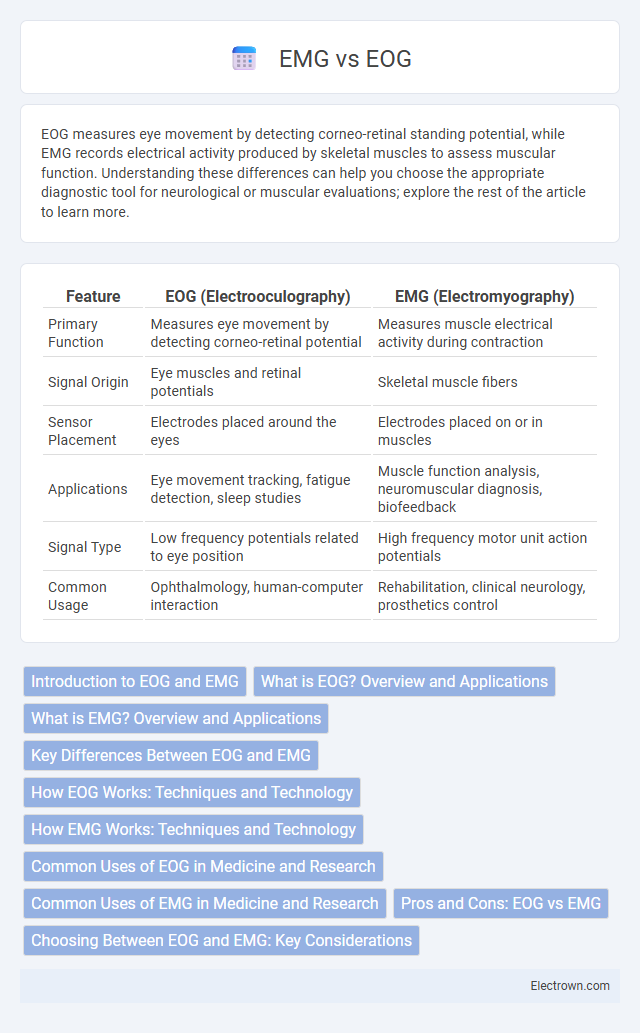
| Feature | EOG (Electrooculography) | EMG (Electromyography) |
|---|---|---|
| Primary Function | Measures eye movement by detecting corneo-retinal potential | Measures muscle electrical activity during contraction |
| Signal Origin | Eye muscles and retinal potentials | Skeletal muscle fibers |
| Sensor Placement | Electrodes placed around the eyes | Electrodes placed on or in muscles |
| Applications | Eye movement tracking, fatigue detection, sleep studies | Muscle function analysis, neuromuscular diagnosis, biofeedback |
| Signal Type | Low frequency potentials related to eye position | High frequency motor unit action potentials |
| Common Usage | Ophthalmology, human-computer interaction | Rehabilitation, clinical neurology, prosthetics control |
Introduction to EOG and EMG
EOG (Electrooculography) measures the electrical potential generated by eye movements, providing valuable data for tracking eye position and diagnosing ophthalmic conditions. EMG (Electromyography) records the electrical activity produced by skeletal muscles, crucial for assessing muscle function and diagnosing neuromuscular disorders. Understanding the fundamentals of EOG and EMG helps you interpret physiological signals critical in medical diagnostics and research.
What is EOG? Overview and Applications
EOG (Electrooculography) measures the resting potential of the retina to track eye movements and detect blink rates by recording electrical signals generated by eye muscles. It is widely used in ophthalmology for diagnosing eye disorders, in sleep studies to monitor REM stages, and in human-computer interaction for eye-tracking applications. Your ability to assess visual attention and ocular motor function improves significantly using EOG technology.
What is EMG? Overview and Applications
EMG (electromyography) measures electrical activity produced by skeletal muscles, providing critical insights into muscle function and neuromuscular health. Common applications of EMG include diagnosing conditions like muscular dystrophy, nerve disorders, and monitoring muscle activation during rehabilitation or athletic training. Your understanding of muscle performance and nerve integrity can be enhanced through EMG, making it a vital tool in clinical and research settings.
Key Differences Between EOG and EMG
Electrooculography (EOG) measures eye movement by detecting corneo-retinal potential changes, while electromyography (EMG) records electrical activity produced by skeletal muscles. EOG primarily monitors ocular motility for applications such as sleep studies and eye-tracking, whereas EMG assesses muscle activation for neuromuscular diagnosis and biofeedback. Understanding the distinction helps you select the appropriate technique for accurate physiological monitoring.
How EOG Works: Techniques and Technology
EOG measures eye movement by detecting voltage changes generated by the corneo-retinal standing potential between the front and back of the human eye using electrodes placed around the eyes. Techniques such as surface electrodes capture these electrical signals to track horizontal and vertical eye movements accurately. This technology enables precise monitoring of eye position and movement, providing valuable data for applications in neurology, sleep studies, and human-computer interaction.
How EMG Works: Techniques and Technology
Electromyography (EMG) measures muscle electrical activity by detecting the electrical potentials generated during muscle contractions using surface or intramuscular electrodes. Techniques include needle EMG, which inserts fine electrodes into muscle tissue for detailed motor unit analysis, and surface EMG, which provides non-invasive monitoring of muscle activation patterns. Advanced EMG technology employs signal amplification, filtering, and real-time data processing to accurately capture and analyze muscle responses for clinical diagnostics and biomechanical research.
Common Uses of EOG in Medicine and Research
Electrooculography (EOG) is primarily used in ophthalmology to assess eye movement disorders, diagnose retinal dysfunction, and evaluate visual system performance. It plays a crucial role in sleep studies by monitoring rapid eye movement (REM) phases and in neuroscience research for analyzing cognitive processes through eye tracking. EOG is also widely applied in brain-computer interface development, enabling communication and control for patients with motor impairments.
Common Uses of EMG in Medicine and Research
Electromyography (EMG) is extensively used in medicine to diagnose neuromuscular disorders such as amyotrophic lateral sclerosis (ALS), myasthenia gravis, and peripheral neuropathies by evaluating muscle electrical activity. In research, EMG is pivotal for studying muscle function, motor control, and rehabilitation outcomes, providing quantitative data on muscle activation patterns during different movements. Compared to electrooculography (EOG), which primarily monitors eye movements, EMG's ability to assess muscle performance makes it essential in clinical neurophysiology and kinesiology research.
Pros and Cons: EOG vs EMG
EOG (Electrooculography) offers precise eye movement tracking with high temporal resolution but can be affected by electrical noise and drift, requiring frequent recalibration. EMG (Electromyography) provides detailed muscle activation data useful for diagnosing neuromuscular disorders but is susceptible to motion artifacts and requires proper electrode placement for accuracy. While EOG excels in eye-related studies, EMG is preferred for muscle function analysis, making their choice dependent on specific diagnostic or research needs.
Choosing Between EOG and EMG: Key Considerations
When choosing between EOG and EMG, consider the specific physiological signals each method detects: EOG measures eye movement through corneo-retinal potential differences, ideal for tracking gaze and blink patterns, while EMG records muscle electrical activity, suited for assessing muscle function and diagnosing neuromuscular disorders. The choice depends on your focus area--EOG is optimal for applications like sleep studies and eye-tracking interfaces, whereas EMG excels in rehabilitation monitoring and biofeedback. Evaluating the required signal type, resolution, and application context ensures accurate data collection tailored to your research or clinical needs.
EOG vs EMG Infographic