MRI provides detailed images of soft tissues using magnetic fields, making it ideal for brain, muscle, and joint evaluations, while CT scans use X-rays for quick imaging of bones, chest, and abdomen, often preferred in emergency settings. Understanding the differences in technology, application, and safety can help you choose the right diagnostic tool; explore the article to learn more about MRI vs CT.
Table of Comparison
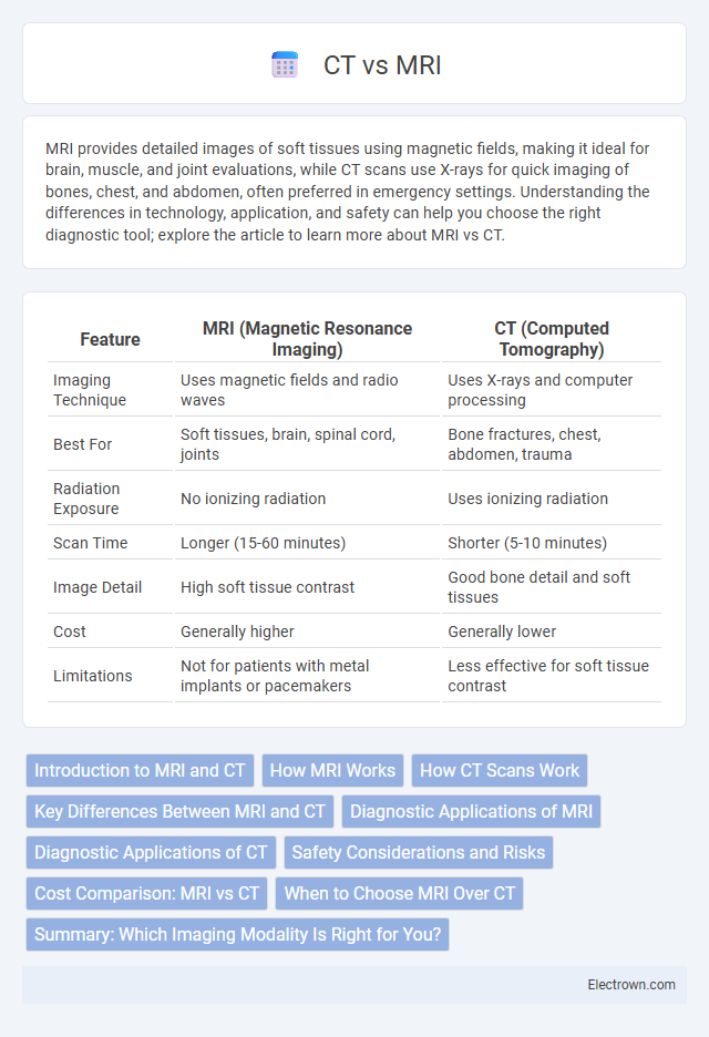
| Feature | MRI (Magnetic Resonance Imaging) | CT (Computed Tomography) |
|---|---|---|
| Imaging Technique | Uses magnetic fields and radio waves | Uses X-rays and computer processing |
| Best For | Soft tissues, brain, spinal cord, joints | Bone fractures, chest, abdomen, trauma |
| Radiation Exposure | No ionizing radiation | Uses ionizing radiation |
| Scan Time | Longer (15-60 minutes) | Shorter (5-10 minutes) |
| Image Detail | High soft tissue contrast | Good bone detail and soft tissues |
| Cost | Generally higher | Generally lower |
| Limitations | Not for patients with metal implants or pacemakers | Less effective for soft tissue contrast |
Introduction to MRI and CT
Magnetic Resonance Imaging (MRI) utilizes strong magnetic fields and radio waves to generate detailed images of soft tissues, organs, and the nervous system without ionizing radiation exposure. Computed Tomography (CT) employs X-ray technology to produce cross-sectional images crucial for diagnosing bone fractures, cancers, and internal bleeding with rapid scan times. Both modalities are essential in medical diagnostics, each offering unique advantages based on tissue contrast, imaging speed, and clinical application.
How MRI Works
Magnetic Resonance Imaging (MRI) operates by using powerful magnetic fields and radio waves to generate detailed images of organs and tissues. Protons in the body's hydrogen atoms align with the magnetic field and emit signals when disrupted by radiofrequency pulses, which are then translated into images by the computer. This technique excels at distinguishing soft tissues without using ionizing radiation, unlike Computed Tomography (CT) scans.
How CT Scans Work
CT scans use X-ray technology to create detailed cross-sectional images by rotating an X-ray tube around the patient and capturing multiple images from different angles. These images are then processed by computer algorithms to generate a 3D representation of the internal structures, highlighting bones, organs, and tissues with high contrast. The rapid acquisition time and high spatial resolution make CT scans particularly effective for detecting fractures, internal bleeding, and tumors.
Key Differences Between MRI and CT
MRI uses strong magnetic fields and radio waves to produce detailed images of soft tissues, making it ideal for brain, muscle, and joint assessments. CT scans rely on X-ray technology to create quick, cross-sectional images, excelling in detecting bone fractures, lung issues, and internal bleeding. Your choice between MRI and CT depends on the area of the body being examined and the specific medical condition requiring diagnosis.
Diagnostic Applications of MRI
MRI excels in diagnosing soft tissue conditions, including brain tumors, spinal cord injuries, and joint abnormalities, offering detailed contrast differentiation not achievable with CT scans. It is particularly effective for detecting neurological disorders, cardiovascular abnormalities, and musculoskeletal injuries due to its ability to produce high-resolution images without ionizing radiation. Your healthcare provider may prefer MRI when precise imaging of soft tissues and functional assessment are critical for accurate diagnosis and treatment planning.
Diagnostic Applications of CT
CT scans provide rapid imaging essential for diagnosing acute conditions such as traumatic brain injuries, pulmonary embolisms, and abdominal emergencies. High-resolution images of bones, blood vessels, and soft tissues enable precise detection of fractures, tumors, and vascular diseases. CT's widespread availability and speed make it invaluable in emergency settings and for guiding interventional procedures.
Safety Considerations and Risks
MRI uses strong magnetic fields and radio waves, avoiding ionizing radiation and minimizing cancer risk, making it safer for repeated imaging and sensitive populations such as pregnant women. CT scans expose patients to ionizing radiation, increasing the risk of radiation-induced malignancies, especially with frequent use or higher doses. Both modalities carry risks related to contrast agents; gadolinium-based contrasts in MRI can lead to nephrogenic systemic fibrosis in patients with kidney impairment, while iodinated contrast in CT may cause allergic reactions or contrast-induced nephropathy.
Cost Comparison: MRI vs CT
MRI scans typically cost significantly more than CT scans due to higher equipment and operational expenses, with MRI procedures averaging between $1,000 and $3,000 compared to CT scans, which range from $300 to $1,500 per session. Insurance coverage varies but often results in higher patient out-of-pocket expenses for MRI, impacting affordability and accessibility. The cost disparity reflects differences in imaging technology complexity, scan duration, and diagnostic detail provided by MRI over CT.
When to Choose MRI Over CT
MRI is preferred over CT when detailed images of soft tissues such as the brain, muscles, ligaments, and spinal cord are required, especially in cases of neurological disorders, tumors, or joint injuries. It provides superior contrast resolution without ionizing radiation, making it ideal for repeated imaging or assessments during pregnancy. MRI is also chosen for evaluating complex conditions like multiple sclerosis, stroke, or abnormalities within the abdomen and pelvis that are less visible on CT scans.
Summary: Which Imaging Modality Is Right for You?
MRI provides superior soft tissue contrast and detailed images, making it ideal for brain, spinal cord, and joint evaluations, while CT scans excel in swiftly detecting bone fractures, acute bleeding, and lung pathology. The choice depends on the clinical context, such as MRI for neurological and musculoskeletal assessments versus CT for trauma and emergency conditions. Considerations also include MRI's longer scan times and contraindications with metal implants versus CT's faster imaging and higher radiation exposure.
MRI vs CT Infographic