Photoplethysmography (PPG) uses light absorption to measure blood volume changes in the microvascular bed, while Impedance Plethysmography (IPG) detects blood flow by measuring electrical impedance variations in body tissues. Understanding the differences between these two techniques can enhance your ability to choose the right method for cardiovascular monitoring, so continue reading to explore their specific applications and advantages.
Table of Comparison
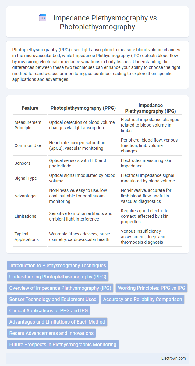
| Feature | Photoplethysmography (PPG) | Impedance Plethysmography (IPG) |
|---|---|---|
| Measurement Principle | Optical detection of blood volume changes via light absorption | Electrical impedance changes related to blood volume in limbs |
| Common Use | Heart rate, oxygen saturation (SpO2), vascular monitoring | Peripheral blood flow, venous function, limb volume changes |
| Sensors | Optical sensors with LED and photodiode | Electrodes measuring skin impedance |
| Signal Type | Optical signal modulated by blood volume | Electrical impedance signal modulated by blood volume |
| Advantages | Non-invasive, easy to use, low cost, suitable for continuous monitoring | Non-invasive, accurate for limb blood flow, useful in vascular diagnostics |
| Limitations | Sensitive to motion artifacts and ambient light interference | Requires good electrode contact; affected by skin properties |
| Typical Applications | Wearable fitness devices, pulse oximetry, cardiovascular health | Venous insufficiency assessment, deep vein thrombosis diagnosis |
Introduction to Plethysmography Techniques
Plethysmography techniques measure changes in volume within an organ or tissue, key for assessing blood flow and vascular health. Photoplethysmography (PPG) uses light absorption variations to detect blood volume changes in the microvascular bed, making it ideal for pulse oximetry and heart rate monitoring. Impedance plethysmography (IPG) measures electrical impedance fluctuations caused by blood volume changes, providing valuable data on limb blood flow and venous function; your choice depends on the monitoring site and clinical application.
Understanding Photoplethysmography (PPG)
Photoplethysmography (PPG) is a non-invasive optical technique that measures blood volume changes in the microvascular bed of tissue, primarily using light absorption variations. It is widely used for monitoring heart rate, blood oxygen saturation, and vascular health by detecting blood flow dynamics through a probe placed on the skin. Compared to Impedance Plethysmography, which measures changes in electrical impedance related to blood volume, PPG offers higher sensitivity to peripheral blood flow and is commonly utilized in wearable health devices.
Overview of Impedance Plethysmography (IPG)
Impedance Plethysmography (IPG) measures changes in electrical impedance caused by blood volume variations in peripheral vessels, providing non-invasive assessment of blood flow and vascular health. Commonly utilized for detecting deep vein thrombosis, arterial occlusions, and evaluating limb edema, IPG offers real-time monitoring with high sensitivity to changes in blood volume and vessel diameter. Its capacity to measure impedance fluctuations contrasts with Photoplethysmography (PPG), which relies on optical absorption changes, making IPG advantageous for evaluating deeper vascular structures and impedance-based volume assessments.
Working Principles: PPG vs IPG
Photoplethysmography (PPG) operates by using a light source and photodetector to measure blood volume changes in microvascular tissue through optical absorption variations, reflecting pulsatile blood flow. Impedance Plethysmography (IPG) measures changes in electrical impedance across a limb or body segment caused by blood volume shifts within the vasculature, using surface electrodes to detect resistance variations. While PPG relies on optical signals and captures arterial pulse waveforms, IPG depends on bioelectrical impedance changes to assess blood flow and volume dynamics.
Sensor Technology and Equipment Used
Photoplethysmography (PPG) utilizes optical sensors, typically infrared or red LEDs and photodetectors, to measure blood volume changes by detecting light absorption variations in the microvascular bed of tissue. Impedance Plethysmography (IPG) relies on electrical sensors, including surface electrodes that apply a small alternating current and measure voltage changes to assess blood flow and volume through tissue impedance. Your choice depends on the need for optical versus electrical measurement methods, with PPG favored for non-invasive, easy-to-use wearable devices and IPG employed in clinical settings requiring precise blood flow analysis.
Accuracy and Reliability Comparison
Photoplethysmography (PPG) provides high accuracy in measuring blood volume changes near the skin surface, making it reliable for heart rate and oxygen saturation monitoring in wearable devices. In contrast, Impedance Plethysmography (IPG) offers greater precision in assessing deeper vascular changes and blood flow dynamics, particularly useful in clinical settings for limb volume and cardiac output measurements. Your choice depends on the specific application, with PPG favoring non-invasive, continuous monitoring accuracy, while IPG excels in detailed vascular assessments requiring reliable impedance data.
Clinical Applications of PPG and IPG
Photoplethysmography (PPG) is extensively used in clinical settings for monitoring cardiovascular health, including heart rate variability, blood oxygen saturation, and peripheral vascular assessments. Impedance Plethysmography (IPG) is primarily applied to evaluate venous insufficiency, arterial occlusion, and blood flow changes in limbs, aiding in diagnosing vascular diseases. Both techniques offer non-invasive, real-time data crucial for managing cardiac and peripheral vascular conditions effectively.
Advantages and Limitations of Each Method
Photoplethysmography (PPG) offers advantages such as non-invasive measurement, high sensitivity to blood volume changes, and ease of use with wearable devices, but it is limited by susceptibility to motion artifacts and poor depth resolution. Impedance plethysmography (IPG) provides accurate assessment of blood flow and volume changes in deeper tissues with better spatial resolution, yet it requires complex electrode placement and can be influenced by skin impedance and external electrical noise. Understanding the limitations and strengths of each method helps you select the appropriate technique for specific clinical or research applications.
Recent Advancements and Innovations
Recent advancements in Photoplethysmography (PPG) include enhanced sensor accuracy through multi-wavelength LEDs and AI-driven signal processing algorithms, improving cardiovascular monitoring precision. In contrast, Impedance Plethysmography (IPG) has seen innovations in wearable electrode materials and integration with portable devices, enabling real-time assessment of limb blood flow and volume changes. Your choice between PPG and IPG benefits from these innovations, as PPG excels in non-invasive pulse and oxygen saturation monitoring, while IPG offers detailed volumetric and vascular resistance data.
Future Prospects in Plethysmographic Monitoring
Future prospects in plethysmographic monitoring emphasize advanced integration of Photoplethysmography (PPG) and Impedance Plethysmography (IPG) to enhance real-time cardiovascular and hemodynamic assessments. Innovations in wearable technology and machine learning algorithms aim to improve signal accuracy, reduce motion artifacts, and enable continuous, non-invasive monitoring for early disease detection. The development of multi-modal devices combining PPG and IPG data promises comprehensive vascular health insights, driving personalized medicine and remote patient management.
Photoplethysmography vs Impedance Plethysmography Infographic