Photoacoustic imaging combines optical and ultrasound techniques to provide high-contrast images of tissue based on light absorption, while ultrasound imaging relies solely on sound waves to visualize structural details. Discover how understanding the differences between these technologies can enhance your diagnostic capabilities by reading the rest of the article.
Table of Comparison
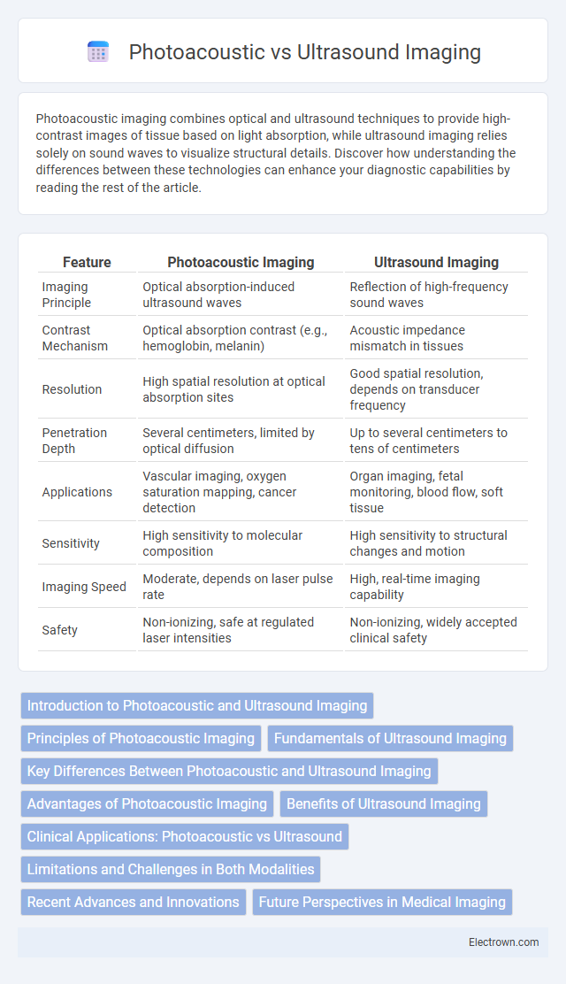
| Feature | Photoacoustic Imaging | Ultrasound Imaging |
|---|---|---|
| Imaging Principle | Optical absorption-induced ultrasound waves | Reflection of high-frequency sound waves |
| Contrast Mechanism | Optical absorption contrast (e.g., hemoglobin, melanin) | Acoustic impedance mismatch in tissues |
| Resolution | High spatial resolution at optical absorption sites | Good spatial resolution, depends on transducer frequency |
| Penetration Depth | Several centimeters, limited by optical diffusion | Up to several centimeters to tens of centimeters |
| Applications | Vascular imaging, oxygen saturation mapping, cancer detection | Organ imaging, fetal monitoring, blood flow, soft tissue |
| Sensitivity | High sensitivity to molecular composition | High sensitivity to structural changes and motion |
| Imaging Speed | Moderate, depends on laser pulse rate | High, real-time imaging capability |
| Safety | Non-ionizing, safe at regulated laser intensities | Non-ionizing, widely accepted clinical safety |
Introduction to Photoacoustic and Ultrasound Imaging
Photoacoustic imaging combines laser-induced ultrasound waves with optical absorption contrast to visualize tissue structures based on their molecular composition, offering high-resolution and functional imaging capabilities. Ultrasound imaging uses high-frequency sound waves to create real-time images of internal organs and soft tissues, relying on acoustic impedance differences for structural visualization. Both modalities complement each other by providing anatomical and molecular information critical for medical diagnostics and research.
Principles of Photoacoustic Imaging
Photoacoustic imaging leverages the photoacoustic effect, where pulsed laser light is absorbed by tissues, causing localized thermal expansion and generating ultrasonic waves detected by ultrasound sensors. This technique combines the high contrast of optical imaging with the spatial resolution of ultrasound, enabling detailed visualization of tissue structure and function. Unlike traditional ultrasound imaging that relies solely on acoustic impedance differences, photoacoustic imaging provides molecular-specific information by detecting optical absorption properties.
Fundamentals of Ultrasound Imaging
Ultrasound imaging utilizes high-frequency sound waves transmitted into the body, where echoes are reflected back from tissue interfaces to create real-time images. The fundamental principles involve piezoelectric transducers that convert electrical signals into sound waves and vice versa, enabling detailed visualization of soft tissues and blood flow. Your diagnostic accuracy benefits from ultrasound's ability to provide continuous imaging without ionizing radiation.
Key Differences Between Photoacoustic and Ultrasound Imaging
Photoacoustic imaging combines laser-induced ultrasound waves generated by optical absorption, providing high-contrast images of tissue oxygenation and molecular composition, while ultrasound imaging relies on sound wave reflections to visualize anatomical structures with real-time detail. Photoacoustic imaging offers superior sensitivity to functional and molecular information, whereas ultrasound excels in structural imaging and depth penetration. Your choice depends on whether you prioritize biochemical insights or anatomical clarity for diagnostic purposes.
Advantages of Photoacoustic Imaging
Photoacoustic imaging offers superior contrast by combining optical absorption properties with ultrasound detection, enabling detailed visualization of vascular structures and oxygenation levels. It provides deeper tissue penetration than pure optical imaging while maintaining high spatial resolution, making it effective for detecting tumors and monitoring physiological changes. This modality also allows non-invasive, real-time imaging without ionizing radiation, enhancing patient safety during diagnostic procedures.
Benefits of Ultrasound Imaging
Ultrasound imaging offers real-time visualization with high spatial resolution and is widely accessible due to its noninvasive, cost-effective nature. It provides excellent soft tissue contrast and allows dynamic assessment of blood flow and organ movement, which is essential for diagnostic and interventional procedures. Your healthcare provider can use ultrasound to quickly obtain detailed anatomical information without exposure to ionizing radiation, ensuring safety and convenience.
Clinical Applications: Photoacoustic vs Ultrasound
Photoacoustic imaging offers superior contrast in visualizing blood vessels and oxygenation levels, making it highly valuable for tumor detection, vascular diseases, and monitoring tissue oxygenation. Ultrasound imaging excel in real-time imaging of soft tissues, guiding biopsies, and assessing organ function such as cardiac and fetal monitoring. Your choice depends on specific clinical needs, with photoacoustic favored for molecular insights and ultrasound for anatomical and functional imaging.
Limitations and Challenges in Both Modalities
Photoacoustic imaging faces challenges such as limited penetration depth due to light scattering in tissues and lower spatial resolution compared to high-frequency ultrasound. Ultrasound imaging is limited by lower contrast resolution when differentiating soft tissues and susceptibility to artifacts caused by bone or gas interference. Both modalities require advanced signal processing and specialized probes to improve image quality and clinical applicability.
Recent Advances and Innovations
Recent advances in photoacoustic imaging leverage enhanced contrast agents and multispectral approaches to enable precise molecular and functional imaging beyond traditional ultrasound capabilities. Innovations in transducer technology and laser integration have improved spatial resolution and imaging depth, facilitating better visualization of vascular structures and tumor identification. These developments position photoacoustic imaging as a complementary modality to ultrasound, integrating structural and functional information for advanced diagnostic applications.
Future Perspectives in Medical Imaging
Photoacoustic imaging combines optical and ultrasound technologies to provide high-contrast, high-resolution images of tissue composition and vascular structures, offering potential for early disease detection and treatment monitoring. Ultrasound imaging remains widely used for real-time visualization of anatomical structures but can benefit from integration with photoacoustic techniques to enhance functional and molecular imaging capabilities. Your future medical diagnostics may rely on hybrid systems that leverage both modalities to improve accuracy, sensitivity, and specificity in complex clinical scenarios.
Photoacoustic vs Ultrasound Imaging Infographic