Chest leads provide detailed information about the horizontal plane of the heart, capturing electrical activity from specific locations on the chest for precise detection of localized abnormalities. Limb leads record electrical signals from the frontal plane, offering a broader view of overall heart rhythm and axis; explore the key differences between chest leads and limb leads in this article to better understand your ECG readings.
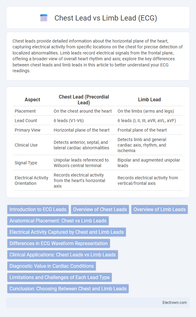
Table of Comparison
| Aspect | Chest Lead (Precordial Lead) | Limb Lead |
|---|---|---|
| Placement | On the chest around the heart | On the limbs (arms and legs) |
| Lead Count | 6 leads (V1-V6) | 6 leads (I, II, III, aVR, aVL, aVF) |
| Primary View | Horizontal plane of the heart | Frontal plane of the heart |
| Clinical Use | Detects anterior, septal, and lateral cardiac abnormalities | Detects limb and general cardiac axis, rhythm, and ischemia |
| Signal Type | Unipolar leads referenced to Wilson's central terminal | Bipolar and augmented unipolar leads |
| Electrical Activity Orientation | Records electrical activity from the heart's horizontal axis | Records electrical activity from vertical/frontal axis |
Introduction to ECG Leads
ECG leads are electrode placements that record electrical activity from different angles of the heart. Chest leads (precordial leads) are positioned on the thorax and provide detailed views of the heart's horizontal plane, especially the anterior and lateral walls. Limb leads, attached to the arms and legs, capture electrical activity in the frontal plane and are essential for assessing overall heart rhythm and axis.
Overview of Chest Leads
Chest leads (precordial leads) in an ECG consist of six electrodes (V1-V6) placed on the thorax, providing a horizontal plane view of the heart's electrical activity. These leads offer detailed information about the anterior, lateral, and septal walls of the left ventricle, crucial for identifying localized cardiac ischemia or infarction. Chest leads detect ventricular depolarization patterns more precisely than limb leads, enhancing the diagnosis of arrhythmias and conduction abnormalities.
Overview of Limb Leads
Limb leads in ECG consist of three bipolar leads (I, II, III) and three augmented unipolar leads (aVR, aVL, aVF) that record electrical activity from the frontal plane of the heart. These leads capture the heart's electrical impulses by measuring voltage differences between electrodes placed on the arms and legs, providing crucial information on cardiac rhythm and axis. Your ECG interpretation benefits from understanding limb leads since they help diagnose arrhythmias, myocardial infarction, and conduction abnormalities.
Anatomical Placement: Chest vs Limb Leads
Chest leads in an ECG are positioned directly on the thorax, providing detailed electrical activity from specific regions of the heart, typically V1 to V6 placed around the sternum and left chest. Limb leads, attached to the arms and legs, including leads I, II, III, aVR, aVL, and aVF, capture the heart's electrical axis in the frontal plane. Understanding the anatomical placement of these leads is essential for accurate ECG interpretation and diagnosing cardiac conditions based on your heart's spatial electrical patterns.
Electrical Activity Captured by Chest and Limb Leads
Chest leads (V1-V6) capture the heart's electrical activity primarily from the horizontal plane, providing detailed information about the anterior, septal, and lateral walls of the ventricles. Limb leads (I, II, III, aVR, aVL, aVF) record electrical signals in the frontal plane, offering insights into the overall heart rhythm and detecting atrial or general ventricular activity. Understanding the differences in electrical activity captured by chest and limb leads helps you accurately interpret cardiac conditions and localize abnormalities within the heart.
Differences in ECG Waveform Representation
Chest leads (precordial leads) provide a horizontal plane view of the heart's electrical activity, capturing waveforms that reflect anterior and lateral ventricular depolarization patterns with higher amplitude and more detailed R wave progression. Limb leads offer a frontal plane perspective, displaying waveforms that emphasize overall heart axis and rhythm by measuring electrical potential differences between the arms and legs. Differences in ECG waveform representation between chest and limb leads are critical for localizing myocardial infarctions and diagnosing conduction abnormalities based on lead-specific variations in wave amplitude, morphology, and polarity.
Clinical Applications: Chest Leads vs Limb Leads
Chest leads (V1-V6) provide detailed information about the anterior, lateral, and septal walls of the heart, making them essential for detecting localized myocardial infarctions and ventricular hypertrophy. Limb leads (I, II, III, aVR, aVL, aVF) offer a broader perspective on the heart's electrical activity, crucial for assessing overall cardiac rhythm, axis deviations, and identifying atrial abnormalities. Combining both lead types enhances diagnostic accuracy in conditions such as ischemia, arrhythmias, and conduction blocks.
Diagnostic Value in Cardiac Conditions
Chest leads (V1-V6) provide detailed information about the anterior, septal, and lateral walls of the heart, making them essential for detecting anterior myocardial infarctions and localized ischemia. Limb leads (I, II, III, aVR, aVL, aVF) offer a broader view of the heart's electrical activity from different angles, crucial for identifying arrhythmias, inferior myocardial infarctions, and axis deviations. Combining chest and limb leads enhances diagnostic accuracy by covering all cardiac regions and improving detection of diverse cardiac conditions.
Limitations and Challenges of Each Lead Type
Chest leads in ECG provide detailed information about the anterior and lateral walls of the heart but are limited by their susceptibility to movement artifacts and difficulty in placement due to anatomical variations. Limb leads offer a broader view of heart activity from different planes but lack the spatial resolution needed to detect localized abnormalities and are prone to interference from limb movement and poor electrode contact. Both lead types require careful interpretation as signal distortion and patient factors can affect diagnostic accuracy.
Conclusion: Choosing Between Chest and Limb Leads
Chest leads provide detailed information about the heart's anterior, septal, and lateral walls, making them essential for detecting localized ischemia or infarction. Limb leads offer a broader view of the heart's electrical activity across the frontal plane, useful for assessing overall rhythm and conduction abnormalities. Selecting between chest and limb leads depends on the clinical requirement for detailed regional analysis versus comprehensive electrical axis evaluation.
Chest Lead vs Limb Lead (ECG) Infographic