Direct ophthalmoscopes provide a magnified, upright view of the retina with a narrow field, ideal for detailed examination of small retinal areas. Indirect ophthalmoscopes offer a wider field of view and a real inverted image, making them better for assessing the peripheral retina; explore the article to discover which ophthalmoscope suits your clinical needs best.
Table of Comparison
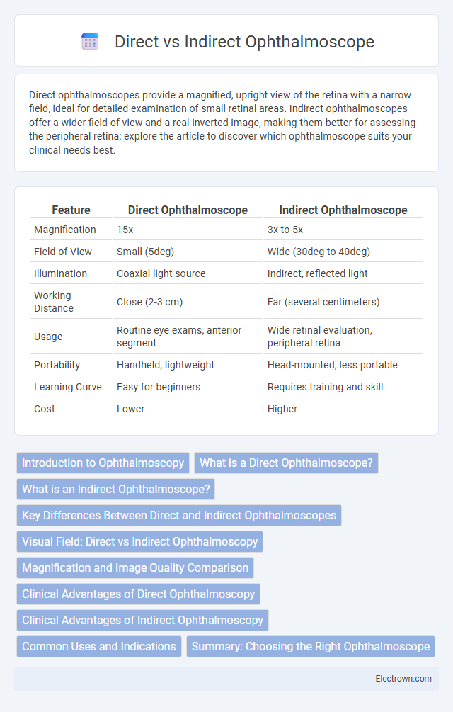
| Feature | Direct Ophthalmoscope | Indirect Ophthalmoscope |
|---|---|---|
| Magnification | 15x | 3x to 5x |
| Field of View | Small (5deg) | Wide (30deg to 40deg) |
| Illumination | Coaxial light source | Indirect, reflected light |
| Working Distance | Close (2-3 cm) | Far (several centimeters) |
| Usage | Routine eye exams, anterior segment | Wide retinal evaluation, peripheral retina |
| Portability | Handheld, lightweight | Head-mounted, less portable |
| Learning Curve | Easy for beginners | Requires training and skill |
| Cost | Lower | Higher |
Introduction to Ophthalmoscopy
Ophthalmoscopy is a crucial diagnostic procedure that allows detailed examination of the eye's internal structures, primarily the retina and optic nerve head. Direct ophthalmoscopes provide a magnified, upright image with a narrow field of view, ideal for detailed inspection of small areas, whereas indirect ophthalmoscopes offer a wider field of view with less magnification, enabling comprehensive assessment of the peripheral retina. Selecting the appropriate ophthalmoscope enhances Your ability to detect and monitor ocular conditions effectively.
What is a Direct Ophthalmoscope?
A direct ophthalmoscope is a handheld device used by eye care professionals to examine the interior structures of the eye, particularly the retina and optic nerve. It provides a magnified, upright, and monocular view, allowing detailed inspection of the fundus through a small pupil. This type of ophthalmoscope is essential for detecting conditions like diabetic retinopathy, glaucoma, and macular degeneration, making it a vital tool in your eye health assessment.
What is an Indirect Ophthalmoscope?
An indirect ophthalmoscope is a specialized optical device used for comprehensive examination of the retina and vitreous body through a wide field of view. It utilizes a binocular headset combined with a condensing lens held close to the patient's eye, allowing detailed assessment of peripheral retinal areas and deeper ocular structures. Your eye care professional relies on this tool for diagnosing retinal detachment, tears, and other posterior segment conditions with enhanced stereoscopic visualization.
Key Differences Between Direct and Indirect Ophthalmoscopes
Direct ophthalmoscopes provide a magnified, upright image of the retina with a narrow field of view, ideal for detailed examination of the optic nerve and macula. Indirect ophthalmoscopes offer a wider field of view with a stereoscopic, inverted image, allowing for comprehensive peripheral retina assessment and detection of retinal detachments. The choice between these instruments depends on diagnostic needs, with direct ophthalmoscopes suited for high-resolution central retina inspection and indirect ophthalmoscopes favored for thorough peripheral retinal evaluation.
Visual Field: Direct vs Indirect Ophthalmoscopy
Direct ophthalmoscopy provides a narrow visual field of approximately 5 degrees, allowing detailed examination of the central retina and optic nerve head. Indirect ophthalmoscopy offers a wider visual field ranging from 20 to 40 degrees, enabling better assessment of the peripheral retina. The broader view in indirect ophthalmoscopy facilitates detection of retinal tears, detachments, and peripheral lesions that may be missed with direct ophthalmoscopy.
Magnification and Image Quality Comparison
Direct ophthalmoscopes provide higher magnification, typically around 15x, allowing for detailed examination of the retina and optic nerve head with a clear, upright image. Indirect ophthalmoscopes offer lower magnification, usually 2x to 5x, but deliver a wider field of view with an inverted, real image, enabling assessment of the peripheral retina. For your diagnostic needs, choosing between magnification and image quality depends on whether you require detailed central retina inspection or broader peripheral visualization.
Clinical Advantages of Direct Ophthalmoscopy
Direct ophthalmoscopy provides high-resolution, magnified images of the retina, enabling detailed examination of the optic disc, macula, and retinal vessels. It offers a small, portable, and easy-to-use device ideal for quick, bedside assessments or primary care settings without requiring pupil dilation. This technique allows for immediate, real-time visualization crucial for diagnosing conditions like diabetic retinopathy and glaucomatous changes efficiently.
Clinical Advantages of Indirect Ophthalmoscopy
Indirect ophthalmoscopy offers a wider field of view and greater depth perception, enabling detailed examination of the retina and peripheral regions that direct ophthalmoscopy might miss. This method is particularly advantageous for diagnosing retinal detachments, diabetic retinopathy, and other peripheral retinal conditions with higher accuracy. Your retinal assessments benefit from enhanced illumination and stereoscopic visualization, crucial for comprehensive ophthalmic evaluations.
Common Uses and Indications
Direct ophthalmoscopes are commonly used for detailed examination of the retina, optic disc, and blood vessels in routine eye check-ups and diagnosing conditions like diabetic retinopathy and glaucoma. Indirect ophthalmoscopes provide a wider field of view and are preferred in assessing peripheral retinal pathology, retinal detachments, and trauma cases. Both instruments are essential in comprehensive ocular evaluations, with direct ophthalmoscopy suited for subtle macular changes and indirect ophthalmoscopy ideal for extensive retinal screening.
Summary: Choosing the Right Ophthalmoscope
Direct ophthalmoscopes provide a single, upright, magnified view of the retina ideal for detailed examination of the central retina and optic disc. Indirect ophthalmoscopes offer a wider, inverted view suitable for peripheral retina evaluation and detecting retinal detachments. Selecting the right ophthalmoscope depends on clinical needs, with direct devices favored for outpatient settings and indirect tools preferred in surgical or emergency ophthalmic care.
Direct vs Indirect Ophthalmoscope Infographic