EOG (Electro-oculography) and ERG (Electroretinography) are specialized diagnostic tests measuring eye function, with EOG assessing the retinal pigment epithelium's health by recording changes in the standing potential of the eye, while ERG evaluates the electrical responses of various retinal cells including photoreceptors and ganglion cells. Understanding the distinctions between EOG and ERG can enhance your grasp of retinal diagnostics; read on to explore their uses, procedures, and clinical significance.
Table of Comparison
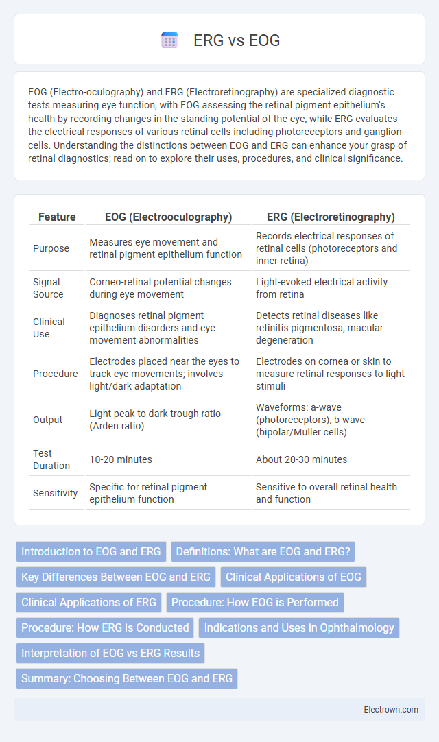
| Feature | EOG (Electrooculography) | ERG (Electroretinography) |
|---|---|---|
| Purpose | Measures eye movement and retinal pigment epithelium function | Records electrical responses of retinal cells (photoreceptors and inner retina) |
| Signal Source | Corneo-retinal potential changes during eye movement | Light-evoked electrical activity from retina |
| Clinical Use | Diagnoses retinal pigment epithelium disorders and eye movement abnormalities | Detects retinal diseases like retinitis pigmentosa, macular degeneration |
| Procedure | Electrodes placed near the eyes to track eye movements; involves light/dark adaptation | Electrodes on cornea or skin to measure retinal responses to light stimuli |
| Output | Light peak to dark trough ratio (Arden ratio) | Waveforms: a-wave (photoreceptors), b-wave (bipolar/Muller cells) |
| Test Duration | 10-20 minutes | About 20-30 minutes |
| Sensitivity | Specific for retinal pigment epithelium function | Sensitive to overall retinal health and function |
Introduction to EOG and ERG
Electrooculography (EOG) measures the electrical potential between the front and back of the eye to assess the function of the retinal pigment epithelium and the outer retina. Electroretinography (ERG) records the electrical responses of various retinal cells, including photoreceptors, bipolar cells, and ganglion cells, to light stimuli, providing detailed information about retinal function. Both EOG and ERG are essential diagnostic tools in ophthalmology, used to evaluate different aspects of retinal health and visual pathway integrity.
Definitions: What are EOG and ERG?
Electrooculography (EOG) measures the resting potential of the retina by recording eye movement potentials, primarily assessing the function of the retinal pigment epithelium and visual cycle. Electroretinography (ERG) records the electrical responses of various retinal cells, including photoreceptors, bipolar cells, and ganglion cells, to light stimuli, providing detailed information about retinal function. Both tests are essential in diagnosing and monitoring ocular and retinal disorders but target different physiological mechanisms within the eye.
Key Differences Between EOG and ERG
Key differences between Electrooculography (EOG) and Electroretinography (ERG) lie in their diagnostic focus and methodology, with EOG measuring the standing potential between the cornea and retina to assess retinal pigment epithelium function, while ERG records electrical responses of various retinal cells to light stimuli, evaluating overall retinal health. EOG primarily detects abnormalities in the retinal pigment epithelium and peripheral retina, commonly used in diagnosing conditions like Best disease, whereas ERG provides detailed information on photoreceptor and inner retinal function, essential for diagnosing retinitis pigmentosa and other retinal dystrophies. The signals obtained are distinct; EOG results are expressed in terms of the Arden ratio, whereas ERG outputs waveform amplitudes and implicit times critical for clinical interpretation.
Clinical Applications of EOG
Electrooculography (EOG) is primarily used to assess the function of the retina and the retinal pigment epithelium by measuring the standing potential between the cornea and the retina, especially in diagnosing conditions like Best disease and other inherited retinal dystrophies. Unlike Electroretinography (ERG), which records electrical responses from photoreceptors and inner retinal cells, EOG evaluates the overall health of the retinal pigment epithelium and can monitor progression in diseases affecting this layer. Your ophthalmologist may recommend EOG testing to complement ERG results and provide a comprehensive evaluation of retinal disorders.
Clinical Applications of ERG
Electroretinography (ERG) is a crucial diagnostic tool in ophthalmology used to assess the electrical responses of various cell types within the retina, including photoreceptors, bipolar cells, and ganglion cells. It aids in the diagnosis and monitoring of retinal disorders such as retinitis pigmentosa, cone-rod dystrophies, and diabetic retinopathy by detecting functional abnormalities before structural changes occur. ERG is also instrumental in evaluating inherited retinal diseases, guiding treatment decisions, and assessing the efficacy of emerging retinal therapies.
Procedure: How EOG is Performed
Electrooculography (EOG) is performed by placing surface electrodes around the eyes to detect resting potential changes associated with eye movement. The patient is asked to perform specific eye movements, such as looking from side to side or up and down, while the electrical activity generated by the retina and eye muscles is recorded. This procedure measures the standing potential between the cornea and retina to evaluate retinal function and diagnose disorders like Best disease or retinitis pigmentosa.
Procedure: How ERG is Conducted
The Electroretinogram (ERG) procedure involves placing a contact lens electrode on the cornea or a skin electrode near the eye to measure electrical responses of retinal cells to light stimuli. Patients experience a series of brief flashes or patterns of light while the retinal response is recorded to assess rod and cone function. This non-invasive test is completed in a controlled, darkened environment to ensure accurate measurement of retinal activity.
Indications and Uses in Ophthalmology
Electrooculography (EOG) is primarily used to assess retinal pigment epithelium function and diagnose conditions such as Best disease and other retinal dystrophies. Electroretinography (ERG) evaluates photoreceptor and inner retinal activity, playing a crucial role in detecting inherited retinal disorders like retinitis pigmentosa and cone-rod dystrophy. Your ophthalmologist may recommend EOG or ERG based on the specific clinical indications to accurately diagnose and monitor retinal health.
Interpretation of EOG vs ERG Results
Interpretation of Electrooculography (EOG) results centers on measuring the standing potential between the cornea and retina, primarily assessing retinal pigment epithelium function. In contrast, Electroretinography (ERG) results provide detailed analysis of the electrical activity generated by retinal cells in response to light stimuli, reflecting photoreceptor and inner retinal function. Clinicians rely on EOG to evaluate conditions like Best disease, while ERG is crucial for diagnosing a wide range of retinal dystrophies and assessing overall retinal health.
Summary: Choosing Between EOG and ERG
EOG (Electrooculography) measures the resting potential of the retina to evaluate eye movement and diagnose retinal pigment epithelium disorders, while ERG (Electroretinography) records electrical responses of various retinal cells to light stimuli, providing detailed retinal function assessment. Your choice depends on the clinical objective: EOG is ideal for detecting retinal pigment epithelium dysfunction, whereas ERG offers comprehensive analysis of photoreceptor and inner retinal activity. Understanding these distinctions ensures accurate diagnosis and tailored treatment in ophthalmologic evaluations.
EOG vs ERG Infographic