Fluoroscopes provide real-time moving images of the internal structures during medical procedures, while radiographs capture static, high-resolution images for diagnostic purposes. Discover the key differences and understand which imaging method best suits your healthcare needs by reading the rest of the article.
Table of Comparison
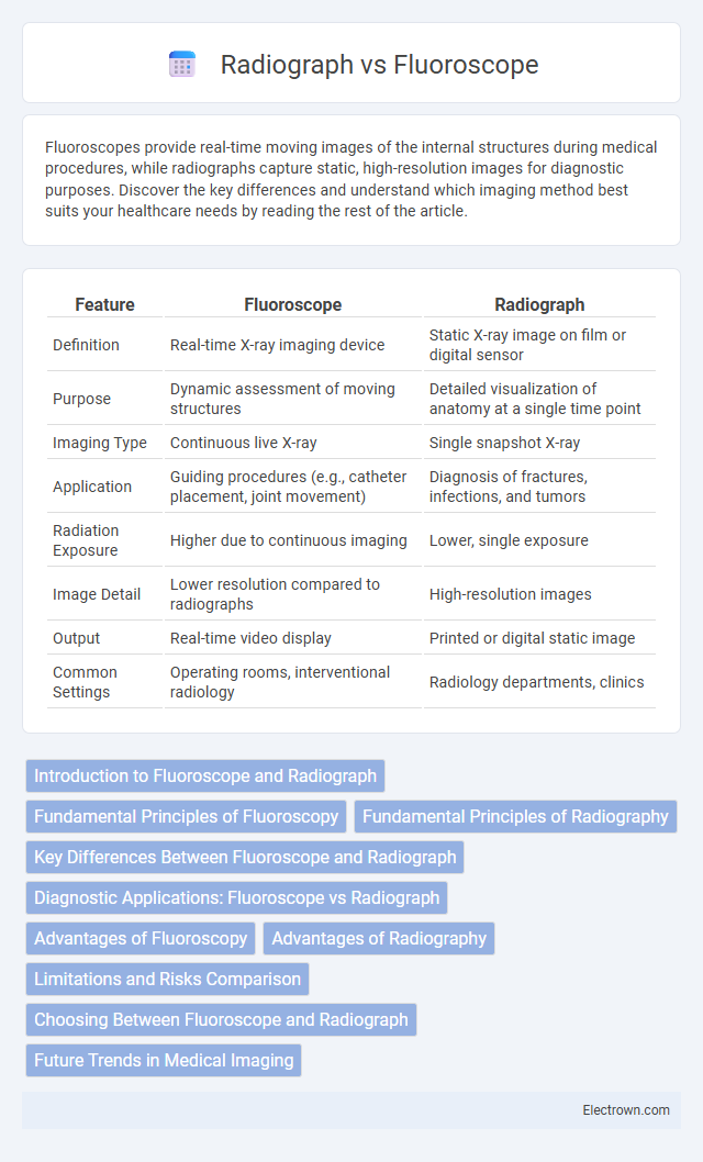
| Feature | Fluoroscope | Radiograph |
|---|---|---|
| Definition | Real-time X-ray imaging device | Static X-ray image on film or digital sensor |
| Purpose | Dynamic assessment of moving structures | Detailed visualization of anatomy at a single time point |
| Imaging Type | Continuous live X-ray | Single snapshot X-ray |
| Application | Guiding procedures (e.g., catheter placement, joint movement) | Diagnosis of fractures, infections, and tumors |
| Radiation Exposure | Higher due to continuous imaging | Lower, single exposure |
| Image Detail | Lower resolution compared to radiographs | High-resolution images |
| Output | Real-time video display | Printed or digital static image |
| Common Settings | Operating rooms, interventional radiology | Radiology departments, clinics |
Introduction to Fluoroscope and Radiograph
Fluoroscopes provide real-time moving images by utilizing continuous X-ray beams, making them essential for dynamic assessments during medical procedures. Radiographs capture static images through a quick burst of X-rays, enabling detailed visualization of bones and tissues for diagnostic purposes. Your choice between a fluoroscope and radiograph depends on whether you need live imaging or a static snapshot to guide diagnosis or treatment.
Fundamental Principles of Fluoroscopy
Fluoroscopy operates on the principle of continuous X-ray beam transmission through the body, producing real-time moving images on a monitor, unlike static radiographs which capture a single snapshot. It utilizes an image intensifier or flat-panel detector to convert X-rays into visible light, enabling dynamic visualization of internal structures, vital for guiding diagnostic and interventional procedures. This real-time imaging capability distinguishes fluoroscopy as an essential tool in fields like cardiology, gastroenterology, and orthopedics.
Fundamental Principles of Radiography
Fluoroscopy utilizes continuous X-ray beams to produce real-time moving images, allowing dynamic assessment of internal structures, whereas radiography captures static images using a single burst of X-rays. Both techniques rely on the differential absorption of X-rays by various tissues, creating contrast based on density and composition. Understanding these fundamental principles helps in selecting the appropriate imaging method for diagnostic clarity and patient safety.
Key Differences Between Fluoroscope and Radiograph
Fluoroscopes provide real-time moving images by continuously emitting X-rays, enabling dynamic observation of internal body structures, unlike radiographs which capture static, single-frame images for detailed examination. Radiographs generally offer higher image resolution and are primarily used for diagnosing bone fractures and evaluating chest conditions, while fluoroscopy is essential for guiding interventional procedures and monitoring organ function. The radiation dose in fluoroscopy is often higher due to prolonged exposure compared to the brief exposure in radiographic imaging.
Diagnostic Applications: Fluoroscope vs Radiograph
Fluoroscopes provide real-time imaging, making them ideal for guiding dynamic diagnostic procedures such as catheter placements, barium swallow studies, and orthopedic assessments. Radiographs capture static, high-resolution images that are essential for diagnosing fractures, infections, and tumors in bones and soft tissues. The choice between fluoroscopy and radiography depends on the need for continuous visualization versus detailed still images in clinical diagnostics.
Advantages of Fluoroscopy
Fluoroscopy offers real-time, dynamic imaging that allows continuous observation of internal structures and functions, making it ideal for guiding diagnostic and interventional procedures. Unlike radiographs, which provide static images, fluoroscopy enables immediate assessment of movement, such as blood flow, gastrointestinal motility, or joint motion, enhancing diagnostic accuracy and treatment precision. Your medical team benefits from fluoroscopy's ability to provide instant feedback during minimally invasive surgeries and catheter placements.
Advantages of Radiography
Radiography offers high-resolution static images that enable detailed examination of bone structures and detection of fractures, making it essential in diagnostic imaging. It provides permanent records that can be easily stored, reviewed, and compared over time, enhancing patient monitoring and treatment planning. Radiographic techniques expose patients to lower radiation doses compared to fluoroscopy, improving safety during routine imaging procedures.
Limitations and Risks Comparison
Fluoroscopy exposes patients to higher doses of ionizing radiation compared to radiographs, increasing the risk of radiation-induced injuries during prolonged procedures. Radiographs provide static images with limited real-time diagnostic capability, potentially missing dynamic abnormalities observable through fluoroscopy. Your choice between these modalities should consider the balance between radiation exposure risks and the specific diagnostic needs of the procedure.
Choosing Between Fluoroscope and Radiograph
Choosing between fluoroscope and radiograph depends on the clinical requirement for real-time imaging or static diagnostic detail. Fluoroscopy provides continuous live X-ray images ideal for guiding procedures such as catheter insertion or gastrointestinal studies, while radiographs offer high-resolution static images essential for detecting fractures or chest abnormalities. Your healthcare provider will decide based on the need for dynamic assessment versus detailed structural evaluation.
Future Trends in Medical Imaging
Fluoroscopes provide real-time moving images, enhancing dynamic assessments during medical procedures, while radiographs offer high-resolution static images essential for detailed anatomical analysis. Emerging trends in medical imaging emphasize integrating artificial intelligence and advanced image processing with both fluoroscopy and radiography, improving diagnostic accuracy and workflow efficiency. Your future healthcare experience may benefit from these innovations through faster diagnostics and personalized imaging protocols tailored to specific clinical needs.
Fluoroscope vs Radiograph Infographic