Surface-enhanced Raman spectroscopy (SERS) significantly amplifies Raman signals by utilizing metal nanostructures, enabling sensitive detection of molecules at very low concentrations. Photoluminescence (PL) spectroscopy analyzes the light emitted by excited materials to provide information about their electronic and structural properties; dive deeper into this article to explore the distinct applications and advantages of each technique for your research needs.
Table of Comparison
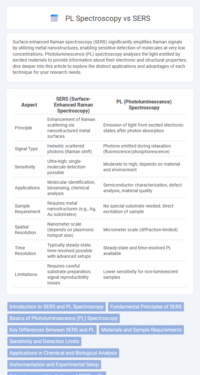
| Aspect | SERS (Surface-Enhanced Raman Spectroscopy) | PL (Photoluminescence) Spectroscopy |
|---|---|---|
| Principle | Enhancement of Raman scattering via nanostructured metal surfaces | Emission of light from excited electronic states after photon absorption |
| Signal Type | Inelastic scattered photons (Raman shift) | Photons emitted during relaxation (fluorescence/phosphorescence) |
| Sensitivity | Ultra-high; single-molecule detection possible | Moderate to high; depends on material and environment |
| Applications | Molecular identification, biosensing, chemical analysis | Semiconductor characterization, defect analysis, material quality |
| Sample Requirement | Requires metal nanostructures (e.g., Ag, Au substrates) | No special substrate needed; direct excitation of sample |
| Spatial Resolution | Nanometer scale (depends on plasmonic hotspot size) | Micrometer scale (diffraction-limited) |
| Time Resolution | Typically steady-state; time-resolved possible with advanced setups | Steady-state and time-resolved PL available |
| Limitations | Requires careful substrate preparation; signal reproducibility issues | Lower sensitivity for non-luminescent samples |
Introduction to SERS and PL Spectroscopy
Surface-Enhanced Raman Scattering (SERS) spectroscopy significantly amplifies Raman signals by utilizing nanostructured metal surfaces, enabling highly sensitive detection of molecular vibrations. Photoluminescence (PL) spectroscopy measures the light emission from excited electrons returning to their ground state, offering insights into electronic and structural properties of materials. Your choice between SERS and PL spectroscopy depends on whether enhanced molecular fingerprinting or detailed electronic state analysis is required for your application.
Fundamental Principles of SERS
Surface-Enhanced Raman Spectroscopy (SERS) amplifies Raman scattering signals by exploiting the localized surface plasmon resonances (LSPRs) of metallic nanostructures such as gold or silver. When molecules are adsorbed onto these nanostructures, electromagnetic enhancement and chemical enhancement mechanisms increase Raman signal intensity by factors up to 10^6 or more, enabling ultra-sensitive molecular detection. Your analytical capabilities benefit from SERS through its ability to provide detailed vibrational fingerprint information with high sensitivity, surpassing the fundamental photoluminescence (PL) spectroscopy principles that rely only on electronic excitations typical of fluorescent processes.
Basics of Photoluminescence (PL) Spectroscopy
Photoluminescence (PL) spectroscopy analyzes the emission of light from a material following photo-excitation, providing insights into electronic and optical properties. It measures the recombination of electron-hole pairs in semiconductors or defects in materials, revealing information about band gaps, impurity states, and exciton dynamics. PL spectroscopy is widely used to characterize nanomaterials, thin films, and quantum dots in optoelectronic and photovoltaic research.
Key Differences Between SERS and PL
Surface-Enhanced Raman Spectroscopy (SERS) amplifies Raman scattering signals using metal nanostructures, providing highly sensitive molecular fingerprint information, while Photoluminescence (PL) spectroscopy measures the light emitted by a sample after excitation, revealing electronic and optical properties. SERS offers enhanced detection of low-concentration analytes with sharp spectral features, whereas PL spectra typically show broader emission bands related to energy band transitions. Your choice depends on whether you need detailed molecular vibrational signatures (SERS) or information on electronic states and recombination processes (PL).
Materials and Sample Requirements
Surface-enhanced Raman spectroscopy (SERS) requires substrates with nanostructured noble metals such as gold, silver, or copper to amplify Raman signals, often necessitating carefully prepared or functionalized surfaces for optimal enhancement. Photoluminescence (PL) spectroscopy depends on materials that exhibit inherent luminescent properties, such as semiconductors, quantum dots, or fluorescent dyes, and typically requires well-prepared samples free of quenching agents. Sample thickness, homogeneity, and background fluorescence critically influence sensitivity and accuracy in both techniques, with SERS favoring thin, adsorbed analytes and PL needing optically transparent samples to prevent signal interference.
Sensitivity and Detection Limits
Surface-Enhanced Raman Spectroscopy (SERS) offers significantly higher sensitivity compared to Photoluminescence (PL) spectroscopy, enabling detection limits down to the single-molecule level due to its enhancement of Raman scattering by metallic nanostructures. PL spectroscopy typically exhibits lower sensitivity because it relies on fluorescence emission, which can be affected by photobleaching and background noise, limiting its detection capabilities to higher analyte concentrations. The ultra-low detection thresholds and rapid signal enhancement of SERS make it a preferred choice for trace-level chemical and biological sensing applications.
Applications in Chemical and Biological Analysis
Surface-enhanced Raman spectroscopy (SERS) offers ultrasensitive detection of trace chemicals and biomolecules by amplifying Raman signals, making it invaluable for identifying pathogens, toxins, and drug molecules in medical diagnostics and environmental monitoring. Photoluminescence (PL) spectroscopy excels in characterizing electronic and structural properties of materials, enabling precise analysis of biological tissues, semiconductor devices, and fluorescent biomarkers. Both techniques complement each other by providing molecular fingerprinting through vibrational modes (SERS) and electronic transitions (PL), enhancing accuracy in chemical and biological analysis.
Instrumentation and Experimental Setup
Surface-Enhanced Raman Spectroscopy (SERS) requires a laser source, Raman spectrometer, and specially prepared metal substrates such as silver or gold nanoparticles to amplify Raman signals. Photoluminescence (PL) spectroscopy involves excitation sources like lasers or lamps, detectors sensitive to emitted light wavelengths, and often cryostats for temperature control to analyze emission properties. Instrumentation in SERS emphasizes enhancement of Raman scattering via nanostructured surfaces, whereas PL setups focus on capturing and characterizing fluorescent or phosphorescent emission from materials.
Advantages and Limitations of SERS vs PL
Surface-enhanced Raman spectroscopy (SERS) offers ultra-sensitive molecular detection by amplifying Raman signals through nanostructured metal surfaces, making it ideal for trace analysis and chemical fingerprinting. Photoluminescence (PL) spectroscopy provides valuable information on electronic and optical properties, but often suffers from lower sensitivity and interference from background fluorescence. Your choice depends on whether you prioritize the high sensitivity and specificity of SERS or the versatile material characterization capabilities of PL, each with distinct limitations such as substrate requirements for SERS and quenching effects in PL.
Future Trends in SERS and PL Spectroscopy
Future trends in SERS (Surface-Enhanced Raman Spectroscopy) emphasize enhanced sensitivity through nanostructured substrates and integration with portable devices for real-time analysis in environmental monitoring and medical diagnostics. PL (Photoluminescence) spectroscopy is advancing with time-resolved techniques and single-molecule detection capabilities, enabling deeper insights in materials science and bioimaging. Both methods are increasingly combined with machine learning algorithms to improve signal interpretation and accelerate high-throughput screening applications.
SERS vs PL spectroscopy Infographic