Scalp EEG offers a non-invasive method to monitor brain activity through electrodes placed on the scalp, while intracranial EEG involves surgically implanted electrodes directly on the brain's surface for more precise signal detection. To understand the advantages and limitations of each technique for your neurological assessment, continue reading the rest of the article.
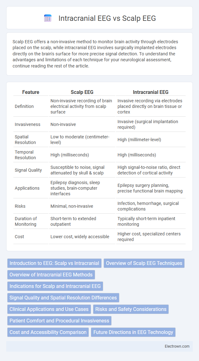
Table of Comparison
| Feature | Scalp EEG | Intracranial EEG |
|---|---|---|
| Definition | Non-invasive recording of brain electrical activity from scalp surface | Invasive recording via electrodes placed directly on brain tissue or cortex |
| Invasiveness | Non-invasive | Invasive (surgical implantation required) |
| Spatial Resolution | Low to moderate (centimeter-level) | High (millimeter-level) |
| Temporal Resolution | High (milliseconds) | High (milliseconds) |
| Signal Quality | Susceptible to noise, signal attenuated by skull & scalp | High signal-to-noise ratio, direct detection of cortical activity |
| Applications | Epilepsy diagnosis, sleep studies, brain-computer interfaces | Epilepsy surgery planning, precise functional brain mapping |
| Risks | Minimal, non-invasive | Infection, hemorrhage, surgical complications |
| Duration of Monitoring | Short-term to extended outpatient | Typically short-term inpatient monitoring |
| Cost | Lower cost, widely accessible | Higher cost, specialized centers required |
Introduction to EEG: Scalp vs Intracranial
Scalp EEG captures electrical brain activity through electrodes placed on the scalp, offering a non-invasive method with broad spatial coverage but limited resolution due to signal attenuation by the skull and scalp. Intracranial EEG involves direct electrode implantation on or within the brain tissue, providing high spatial and temporal resolution critical for precise localization of epileptogenic zones. The choice between scalp and intracranial EEG depends on clinical needs, balancing invasiveness against the accuracy of neural signal detection.
Overview of Scalp EEG Techniques
Scalp EEG techniques involve non-invasive placement of electrodes on the scalp to record electrical activity generated by cortical neurons, providing valuable insight into brain function with minimal risk. Common configurations include the 10-20 system, which ensures standardized electrode placement for consistent data collection and clinical interpretation. These methods excel at detecting generalized and focal epileptiform discharges, making them essential tools for diagnosing epilepsy, monitoring sleep patterns, and assessing brain injuries.
Overview of Intracranial EEG Methods
Intracranial EEG (iEEG) involves direct recording of electrical activity from the brain surface or within brain tissue using implanted electrodes, providing higher spatial resolution and signal fidelity than scalp EEG. Common methods include subdural grid electrodes, depth electrodes, and stereo-EEG (SEEG), each offering precise localization for epilepsy surgery planning and investigating cortical functions. This invasive approach allows detection of high-frequency oscillations and ictal onset zones inaccessible to scalp EEG, crucial for tailoring patient-specific interventions.
Indications for Scalp and Intracranial EEG
Scalp EEG is primarily indicated for non-invasive assessment of generalized and focal epilepsy, sleep disorders, and encephalopathies, offering surface-level brain activity monitoring. Intracranial EEG is reserved for presurgical evaluation of refractory focal epilepsy when scalp EEG findings are inconclusive or insufficient for precise localization of epileptogenic zones. The choice depends on the clinical need for spatial resolution, diagnostic clarity, and risk-benefit analysis in epilepsy treatment planning.
Signal Quality and Spatial Resolution Differences
Intracranial EEG offers superior signal quality and spatial resolution compared to scalp EEG by directly recording electrical activity from the brain surface or depth, minimizing signal attenuation and artifacts caused by the skull and scalp. Scalp EEG captures broader cortical activity with lower spatial resolution due to signal diffusion and interference, limiting precise localization of epileptogenic zones. The enhanced precision of intracranial EEG enables accurate mapping of epileptic foci crucial for surgical planning in refractory epilepsy cases.
Clinical Applications and Use Cases
Scalp EEG is widely used for non-invasive monitoring of brain activity in routine clinical diagnosis, especially for epilepsy, sleep disorders, and encephalopathies due to its ability to capture generalized cortical activity. Intracranial EEG, offering direct brain surface or depth recordings, is essential for pre-surgical evaluation in refractory epilepsy patients to precisely localize seizure foci and guide surgical intervention. Understanding these clinical applications helps you select the appropriate EEG modality for accurate diagnosis and effective treatment planning.
Risks and Safety Considerations
Scalp EEG offers a non-invasive method with minimal risk, primarily causing mild skin irritation or discomfort from electrode placement. Intracranial EEG involves surgical implantation, carrying risks such as infection, bleeding, and potential neurological damage due to its invasive nature. Your healthcare provider will weigh these safety considerations carefully to determine the most appropriate and safest option based on your clinical needs.
Patient Comfort and Procedural Invasiveness
Scalp EEG offers greater patient comfort due to its non-invasive application, involving electrodes placed on the scalp without surgical intervention. Intracranial EEG requires invasive procedures, such as craniotomy or stereotactic electrode implantation, which can cause discomfort and increased risk of complications. The procedural invasiveness of intracranial EEG limits its use to cases where detailed cortical activity monitoring is essential, despite lower patient comfort compared to scalp EEG.
Cost and Accessibility Comparison
Scalp EEG offers a cost-effective and widely accessible method for monitoring brain activity, making it suitable for routine diagnostic use across most healthcare settings. Intracranial EEG, while providing higher spatial resolution, involves significantly higher costs due to surgical implantation and specialized monitoring equipment, limiting its availability to specialized centers. Your decision between the two should consider budget constraints and the necessity for detailed neural data depending on the clinical scenario.
Future Directions in EEG Technology
Future directions in EEG technology emphasize enhancing the resolution and accuracy of both Scalp and Intracranial EEG by integrating advanced signal processing algorithms and machine learning techniques. Innovations aim to improve non-invasive scalp EEG's spatial resolution, approaching the precision of intracranial EEG, while minimizing patient risk. Your ability to monitor and interpret brain activity will benefit from wearable and wireless EEG devices that facilitate continuous, real-time data collection and analysis.
Scalp EEG vs Intracranial EEG Infographic