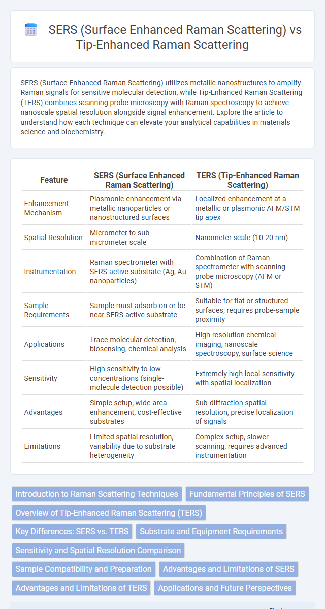
SERS (Surface Enhanced Raman Scattering) utilizes metallic nanostructures to amplify Raman signals for sensitive molecular detection, while Tip-Enhanced Raman Scattering (TERS) combines scanning probe microscopy with Raman spectroscopy to achieve nanoscale spatial resolution alongside signal enhancement. Explore the article to understand how each technique can elevate your analytical capabilities in materials science and biochemistry.
Table of Comparison
| Feature | SERS (Surface Enhanced Raman Scattering) | TERS (Tip-Enhanced Raman Scattering) |
|---|---|---|
| Enhancement Mechanism | Plasmonic enhancement via metallic nanoparticles or nanostructured surfaces | Localized enhancement at a metallic or plasmonic AFM/STM tip apex |
| Spatial Resolution | Micrometer to sub-micrometer scale | Nanometer scale (10-20 nm) |
| Instrumentation | Raman spectrometer with SERS-active substrate (Ag, Au nanoparticles) | Combination of Raman spectrometer with scanning probe microscopy (AFM or STM) |
| Sample Requirements | Sample must adsorb on or be near SERS-active substrate | Suitable for flat or structured surfaces; requires probe-sample proximity |
| Applications | Trace molecular detection, biosensing, chemical analysis | High-resolution chemical imaging, nanoscale spectroscopy, surface science |
| Sensitivity | High sensitivity to low concentrations (single-molecule detection possible) | Extremely high local sensitivity with spatial localization |
| Advantages | Simple setup, wide-area enhancement, cost-effective substrates | Sub-diffraction spatial resolution, precise localization of signals |
| Limitations | Limited spatial resolution, variability due to substrate heterogeneity | Complex setup, slower scanning, requires advanced instrumentation |
Introduction to Raman Scattering Techniques
Surface-enhanced Raman scattering (SERS) significantly amplifies Raman signals by leveraging plasmonic nanostructures, achieving sensitivity down to single-molecule detection. Tip-enhanced Raman scattering (TERS) combines atomic force microscopy (AFM) or scanning tunneling microscopy (STM) with localized surface plasmon resonance to provide spatial resolution at the nanometer scale while enhancing Raman scattering. Both techniques enhance traditional Raman spectroscopy but differ in their enhancement mechanisms and spatial resolution capabilities, enabling advanced chemical and structural analysis at varying scales.
Fundamental Principles of SERS
Surface-enhanced Raman scattering (SERS) enhances Raman signals by exploiting localized surface plasmon resonances on nanoscale metallic structures, typically silver or gold nanoparticles, which amplify the electromagnetic fields near the surface. This amplification occurs when incident light excites collective electron oscillations, dramatically increasing the Raman scattering cross-section of molecules adsorbed on or near the metallic surface. SERS relies primarily on electromagnetic enhancement combined with chemical enhancement mechanisms originating from charge transfer interactions between the metal and the analyte.
Overview of Tip-Enhanced Raman Scattering (TERS)
Tip-Enhanced Raman Scattering (TERS) combines scanning probe microscopy with Raman spectroscopy, enabling molecular-scale chemical imaging with enhanced spatial resolution down to a few nanometers. Unlike surface-enhanced Raman scattering (SERS), which relies on plasmonic substrates to amplify Raman signals over larger areas, TERS uses a sharp metallic tip to localize the electromagnetic enhancement precisely at the tip apex, providing superior sensitivity and spatial resolution. Your analysis benefits from TERS when investigating nanoscale surface chemistry and molecular structures with high specificity and minimal sample preparation.
Key Differences: SERS vs. TERS
SERS (Surface Enhanced Raman Scattering) enhances Raman signals by using nanostructured metallic surfaces like gold or silver, while TERS (Tip-Enhanced Raman Scattering) achieves localized enhancement with a metallic or plasmonic AFM/STM tip for nanoscale spatial resolution. SERS provides signal amplification over larger surface areas, whereas TERS enables Raman spectroscopy with spatial resolution down to 10-20 nanometers, allowing molecular level imaging. The main difference lies in TERS's capability for site-specific enhancement, combining tip-induced field concentration with scanning probe microscopy, which SERS lacks.
Substrate and Equipment Requirements
Surface-enhanced Raman scattering (SERS) relies on metallic nanostructured substrates, commonly silver or gold nanoparticles, to amplify Raman signals through localized surface plasmon resonance, requiring precise nanofabrication or colloidal synthesis for effective enhancement. Tip-enhanced Raman scattering (TERS) combines scanning probe microscopy with Raman spectroscopy, employing a sharp metallic tip, often gold or silver-coated, to create a highly localized enhancement at the nanoscale, demanding sophisticated combined instrumentation with atomic force or scanning tunneling microscopes. Your choice between SERS and TERS depends on required spatial resolution and available equipment, as SERS substrates are generally simpler and more scalable, while TERS offers superior nanoscale specificity but requires complex and costly apparatus.
Sensitivity and Spatial Resolution Comparison
Surface-enhanced Raman scattering (SERS) offers high sensitivity by amplifying Raman signals via plasmonic nanostructures, achieving detection limits down to single molecules under optimal conditions. Tip-enhanced Raman scattering (TERS) surpasses SERS in spatial resolution, reaching sub-10-nanometer levels by combining scanning probe microscopy with localized plasmonic enhancement at the tip apex. While SERS exhibits broad-area enhancement suited for ensemble measurements, TERS provides nanoscale chemical imaging with superior spatial precision but typically lower overall signal intensity compared to SERS.
Sample Compatibility and Preparation
Surface Enhanced Raman Scattering (SERS) requires the sample to be in close proximity to or adsorbed onto metallic nanostructures, often necessitating careful preparation such as nanoparticle synthesis or substrate functionalization to ensure effective plasmonic enhancement. Tip-Enhanced Raman Scattering (TERS) allows direct analysis of samples on flat or rough surfaces without extensive chemical preparation, as the enhancement is localized at the apex of a metallic tip, offering more flexibility in sample compatibility. Your choice between SERS and TERS should consider the nature of the sample and preparation complexity, with TERS being advantageous for delicate or heterogeneous materials.
Advantages and Limitations of SERS
SERS (Surface Enhanced Raman Scattering) offers ultra-sensitive detection by amplifying Raman signals using metallic nanostructures, enabling trace-level chemical and biological analysis. Its advantages include high enhancement factors, rapid response, and ease of use for large-area sensing, but limitations involve reproducibility challenges and dependence on the quality of the nanostructured substrate. Unlike Tip-Enhanced Raman Scattering (TERS), SERS provides broader sample coverage but lacks the nanoscale spatial resolution and localized enhancement achievable with TERS.
Advantages and Limitations of TERS
Tip-enhanced Raman scattering (TERS) offers unparalleled spatial resolution down to the nanoscale, enabling detailed chemical and structural analysis beyond the diffraction limit of light. Unlike surface-enhanced Raman scattering (SERS), which relies on rough metallic surfaces or nanoparticles for signal enhancement, TERS uses a sharp metallic tip to provide localized and highly controlled enhancement, making it ideal for studying individual molecules or nanoscale features. The main limitations of TERS include its complex experimental setup, sensitivity to tip contamination or damage, and typically slower acquisition times compared to SERS, which can restrict its practical applicability in high-throughput analyses.
Applications and Future Perspectives
Surface-enhanced Raman scattering (SERS) is widely used for rapid chemical sensing, biosensing, and environmental monitoring due to its high sensitivity and ability to detect trace molecules on metal nanostructures. Tip-enhanced Raman scattering (TERS) offers nanoscale spatial resolution, enabling detailed surface characterization in materials science, catalysis, and biological studies by combining scanning probe microscopy with Raman spectroscopy. Your choice between SERS and TERS depends on whether you prioritize sensitivity for bulk analysis or spatial resolution for nanoscale imaging, with future advancements expected to enhance their integration in real-time diagnostics and advanced nanotechnology applications.
SERS (Surface enhanced Raman scattering) vs Tip-enhanced Raman scattering Infographic